Astrocytes may be a key player in the brain’s ability to process external and internal information simultaneously, according to a new study.
Long thought of as “brain glue,” the star-shaped cells called astrocytes are members of a family of cells found in the central nervous system called glial that help regulate blood flow and synaptic activity, keep neurons healthy, and play an important role in breathing.
Despite this growing appreciation for astrocytes, much remains unknown about the role these cells play in helping neurons and the brain process information.
“We believe astrocytes can add a new dimension to our understanding of how external and internal information is merged in the brain,” says Nathan Smith, associate professor of neuroscience at the Del Monte Institute for Neuroscience at the University of Rochester.
“More research on these cells is necessary to understand their role in the process that allows a person to have an appropriate behavioral response and also the ability to create a relevant memory to guide future behavior.”
The way our body integrates external with internal information is essential to survival. When something goes awry in these processes, behavioral or psychiatric symptoms may emerge.
Smith and coauthors point to evidence that astrocytes may play a key role in this process. Previous research has shown astrocytes sense the moment neurons send a message and can simultaneously sense sensory inputs. These external signals could come from various senses such as sight or smell.
Astrocytes respond to this influx of information by modifying their calcium Ca2+ signaling directed towards neurons, providing them with the most suitable information to react to the stimuli.
The authors hypothesize that this astrocytic Ca2+ signaling may be an underlying factor in how neurons communicate and what may happen when a signal is disrupted. But much is still unknown in how astrocytes and neuromodulators, the signals sent between neurons, work together.
“Astrocytes are an often-overlooked type of brain cell in systems neuroscience,” Smith says. “We believe dysfunctional astrocytic calcium signaling could be an underlying factor in disorders characterized by disrupted sensory processing, like Alzheimer’s and autism spectrum disorder.”
Smith has spent his career studying astrocytes. As a graduate student at the University of Rochester School of Medicine and Dentistry, Smith was part of the team who discovered an expanded role for astrocytes. Apart from absorbing excess potassium, astrocytes themselves could cause potassium levels around the neuron to drop, halting neuronal signaling. This research showed, for the first time, that astrocytes did more than tend to neurons, they also could influence the actions of neurons.
“I think once we understand how astrocytes integrate external information from these different internal states, we can better understand certain neurological diseases. Understanding their role more fully will help propel the future possibility of targeting astrocytes in neurological disease,” Smith says.
The communication between neurons and astrocytes is far more complicated than previously thought. Evidence suggests that astrocytes can sense and react to change—a process that is important for behavioral shifts and memory formation.
The study authors believe discovering more about astrocytes will lead to a better understanding of cognitive function and lead to advances in treatment and care.
The study appears in Trends in Neuroscience.
Additional coauthors are from the University of Copenhagen.
The National Institutes of Health, the National Science Foundation, the European Union under the Marie Skłodowska-Curie Fellowship, the ONO Rising Star Fellowship, the Lundbeck Foundation Experiment Grant, and the Novo Nordisk Foundation supported the work.
Source: University of Rochester
The post Star-shaped cells may play role in how your brain merges info appeared first on Futurity.

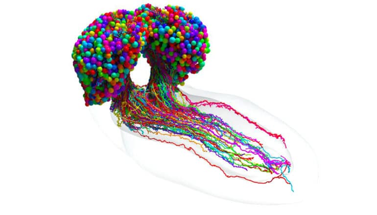
The most advanced brain map to date, that of an insect—brings scientists closer to true understanding of the mechanism of thought.
The researchers produced a breathtakingly detailed diagram tracing every neural connection in the brain of a larval fruit fly, an archetypal scientific model with brains comparable to humans.
The work, likely to underpin future brain research and inspire new machine learning architectures, appears in the journal Science.
“Everything has been working up to this.”
“If we want to understand who we are and how we think, part of that is understanding the mechanism of thought,” says senior author Joshua T. Vogelstein, a biomedical engineer at Johns Hopkins University who specializes in data-driven projects including connectomics, the study of nervous system connections. “And the key to that is knowing how neurons connect with each other.”
The first attempt at mapping a brain—a 14-year study of the roundworm begun in the 1970s, resulted in a partial map and a Nobel Prize. Since then, partial connectomes have been mapped in many systems, including flies, mice, and even humans, but these reconstructions typically represent only a tiny fraction of the total brain.
Comprehensive connectomes have only been generated for several small species with a few hundred to a few thousand neurons in their bodies: a roundworm, a larval sea squirt, and a larval marine annelid worm.
This team’s connectome of a baby fruit fly, Drosophila melanogaster larva, is the most complete as well as the most expansive map of an entire insect brain ever completed. It includes 3,016 neurons and every connection between them: 548,000.
“It’s been 50 years and this is the first brain connectome. It’s a flag in the sand that we can do this,” Vogelstein says. “Everything has been working up to this.”
Mapping whole brains is difficult and extremely time-consuming, even with the best modern technology. Getting a complete cellular-level picture of a brain requires slicing the brain into hundreds or thousands of individual tissue samples, all of which have to be imaged with electron microscopes before the painstaking process of reconstructing all those pieces, neuron by neuron, into a full, accurate portrait of a brain.
It took more than a decade to do that with the baby fruit fly. The brain of a mouse is estimated to be a million times larger than that of a baby fruit fly, meaning the chance of mapping anything close to a human brain isn’t likely in the near future, maybe not even in our lifetimes.
The team purposely chose the fruit fly larva because, for an insect, the species shares much of its fundamental biology with humans, including a comparable genetic foundation. It also has rich learning and decision-making behaviors, making it a useful model organism in neuroscience. And for practical purposes, its relatively compact brain can be imaged and its circuits reconstructed within a reasonable time frame.
Even so, the work took the University of Cambridge and Johns Hopkins 12 years. The imaging alone took about a day per neuron.
Cambridge researchers created the high-resolution images of the brain and manually studied them to find individual neurons, rigorously tracing each one and linking their synaptic connections.
Cambridge handed off the data to Johns Hopkins, where the team spent more than three years using original code they created to analyze the brain’s connectivity. The Johns Hopkins team developed techniques to find groups of neurons based on shared connectivity patterns, and then analyzed how information could propagate through the brain.
In the end, the full team charted every neuron and every connection, and categorized each neuron by the role it plays in the brain. They found that the brain’s busiest circuits were those that led to and away from neurons of the learning center.
The methods the researchers developed are applicable to any brain connection project, and their code is available to whoever attempts to map an even larger animal brain, Vogelstein says, adding that despite the challenges, scientists are expected to take on the mouse, possibly within the next decade.
Other teams are already working on a map of the adult fruit fly brain. Co-first author Benjamin Pedigo, a Johns Hopkins doctoral candidate in biomedical engineering, expects the team’s code could help reveal important comparisons between connections in the adult and larval brain. As connectomes are generated for more larva and from other related species, Pedigo expects their analysis techniques could lead to better understanding of variations in brain wiring.
The fruit fly larva work showed circuit features that were strikingly reminiscent of prominent and powerful machine learning architectures. The team expects continued study will reveal even more computational principles and potentially inspire new artificial intelligence systems.
“What we learned about code for fruit flies will have implications for the code for humans,” Vogelstein says. “That’s what we want to understand—how to write a program that leads to a human brain network.”
Source: Johns Hopkins University
The post First map of insect brain could shed light on thinking appeared first on Futurity.