On this episode of the Big Brains podcast, a scholar explains the neuroscience of how listening to and playing music builds our mind.
Music plays an important role in all of our lives. But listening to music or playing an instrument is more than just a creative outlet or hobby—it’s also scientifically good for us. Research shows that music can stimulate new connections in our brains; keeping our cognitive abilities sharp and our memories alive.
In a new book, Every Brain Needs Music: The Neuroscience of Making and Listening to Music (Columbia University Press, 2023), Larry Sherman explores why we all need music for our mental well-being—and how it can even help us later in life.
Sherman is a professor of neuroscience at Oregon Health & Science University.
Listen to the episode below:
Read the transcript to the episode. Subscribe to Big Brains on Apple Podcasts, Stitcher, and Spotify.
Source: University of Chicago
The post How music benefits your brain appeared first on Futurity.
New research clarifies how the brain goes to great lengths to process and remember everyday events.
Researchers used functional MRI scanners to monitor the brains of subjects watching short videos of scenes that could have come from real life. These included people working on laptops in a cafe or shopping in a grocery store.
“They were very ordinary scenes,” says Zachariah Reagh, an assistant professor of psychological and brain sciences at Washington University in St. Louis. “No car chases or anything.”
The research subjects then immediately described the scenes with as much detail as they could muster. The mundane snippets led to intriguing findings, including that different parts of the brain worked together to understand and remember a situation.
Networks in the front part of the temporal lobe, a region of the brain long known to play an important role in memory, focused on the subject regardless of their surroundings. But the posterior medial network, which involves the parietal lobe toward the back of the brain, paid more attention to the environment. Those networks then sent information to the hippocampus, Reagh explains, which combined the signals to create a cohesive scene.
Researchers had previously used very simple objects and scenarios—such as a picture of an apple on a beach—to study the different building blocks of memories, Reagh says. But life isn’t so simple, he says. “I wondered if anyone had done these types of studies with dynamic real-word situations and, shockingly, the answer was no.”
The new study in Nature Communications suggests that the brain makes mental sketches of people that can be transposed from one location to another, much like an animator can copy and paste a character into different scenes. “It may not seem intuitive that your brain can create a sketch of a family member that it moves from place to place, but it’s very efficient,” he says.
Some subjects could recall the scenes in the café and grocery store more completely and accurately than others. Reagh and coauthor Charan Ranganath of the University of California, Davis, found that those with the clearest memories used the same neural patterns when recalling scenes that they used while watching the clips. “The more you can bring those patterns back online while describing an event, the better your overall memory,” he says.
At this time, Reagh says, it’s unclear why some people seem more adept than others at reproducing the thought patterns needed to access memory. But it’s clear that many things can get in the way. “A lot can go wrong when you try to retrieve a memory,” he says.
Even memories that seem crisp and vivid may not actually reflect reality. “I tell my students that your memory is not a video camera. It doesn’t give you a perfect representation of what happened. Your brain is telling you a story,” he says.
In future, Reagh plans to study the brain activity and memory of people watching more complicated stories.
Source: Washington University in St. Louis
The post Mundane scenes clarify how brain makes memories appeared first on Futurity.
Astrocytes may be a key player in the brain’s ability to process external and internal information simultaneously, according to a new study.
Long thought of as “brain glue,” the star-shaped cells called astrocytes are members of a family of cells found in the central nervous system called glial that help regulate blood flow and synaptic activity, keep neurons healthy, and play an important role in breathing.
Despite this growing appreciation for astrocytes, much remains unknown about the role these cells play in helping neurons and the brain process information.
“We believe astrocytes can add a new dimension to our understanding of how external and internal information is merged in the brain,” says Nathan Smith, associate professor of neuroscience at the Del Monte Institute for Neuroscience at the University of Rochester.
“More research on these cells is necessary to understand their role in the process that allows a person to have an appropriate behavioral response and also the ability to create a relevant memory to guide future behavior.”
The way our body integrates external with internal information is essential to survival. When something goes awry in these processes, behavioral or psychiatric symptoms may emerge.
Smith and coauthors point to evidence that astrocytes may play a key role in this process. Previous research has shown astrocytes sense the moment neurons send a message and can simultaneously sense sensory inputs. These external signals could come from various senses such as sight or smell.
Astrocytes respond to this influx of information by modifying their calcium Ca2+ signaling directed towards neurons, providing them with the most suitable information to react to the stimuli.
The authors hypothesize that this astrocytic Ca2+ signaling may be an underlying factor in how neurons communicate and what may happen when a signal is disrupted. But much is still unknown in how astrocytes and neuromodulators, the signals sent between neurons, work together.
“Astrocytes are an often-overlooked type of brain cell in systems neuroscience,” Smith says. “We believe dysfunctional astrocytic calcium signaling could be an underlying factor in disorders characterized by disrupted sensory processing, like Alzheimer’s and autism spectrum disorder.”
Smith has spent his career studying astrocytes. As a graduate student at the University of Rochester School of Medicine and Dentistry, Smith was part of the team who discovered an expanded role for astrocytes. Apart from absorbing excess potassium, astrocytes themselves could cause potassium levels around the neuron to drop, halting neuronal signaling. This research showed, for the first time, that astrocytes did more than tend to neurons, they also could influence the actions of neurons.
“I think once we understand how astrocytes integrate external information from these different internal states, we can better understand certain neurological diseases. Understanding their role more fully will help propel the future possibility of targeting astrocytes in neurological disease,” Smith says.
The communication between neurons and astrocytes is far more complicated than previously thought. Evidence suggests that astrocytes can sense and react to change—a process that is important for behavioral shifts and memory formation.
The study authors believe discovering more about astrocytes will lead to a better understanding of cognitive function and lead to advances in treatment and care.
The study appears in Trends in Neuroscience.
Additional coauthors are from the University of Copenhagen.
The National Institutes of Health, the National Science Foundation, the European Union under the Marie Skłodowska-Curie Fellowship, the ONO Rising Star Fellowship, the Lundbeck Foundation Experiment Grant, and the Novo Nordisk Foundation supported the work.
Source: University of Rochester
The post Star-shaped cells may play role in how your brain merges info appeared first on Futurity.
Scientists have gained new insights into how the brain’s internal compass gives us a sense of direction.
The findings shed light on how the brain orients itself in changing environments—and even the processes that can go wrong with degenerative diseases like dementia that leave people feeling lost and confused.
“Neuroscience research has witnessed a technology revolution in the last decade allowing us to ask and answer questions that could only be dreamed of just years ago,” says Mark Brandon, an associate professor of psychiatry at McGill University and researcher at the Douglas Research Centre who co-led the work with Zaki Ajabi, a former student at McGill University and now a postdoctoral research fellow at Harvard University.
To understand how visual information affects the brain’s internal compass, the researchers exposed mice to a disorienting virtual world while recording the brain’s neural activity. The team recorded the brain’s internal compass with unprecedented precision using the latest advances in neuronal recording technology.
This ability to accurately decode the animal’s internal head direction allowed the researchers to explore how the head-direction cells, which make up the brain’s internal compass, support the brain’s ability to reorient itself in changing surroundings.
Specifically, the research team identified a phenomenon they call “network gain” that allowed the brain’s internal compass to reorient after the mice were disoriented.
“It’s as if the brain has a mechanism to implement a ‘reset button’ allowing for rapid reorientation of its internal compass in confusing situations,” says Ajabi.
Although researchers exposed the animals in this study to unnatural visual experiences, the authors argue that such scenarios are already relevant to the modern human experience, especially with the rapid spread of virtual reality technology.
These findings “may eventually explain how virtual reality systems can easily take control over our sense of orientation,” adds Ajabi.
The results inspired the research team to develop new models to better understand the underlying mechanisms.
“This work is a beautiful example of how experimental and computational approaches together can advance our understanding of brain activity that drives behavior,” says coauthor Xue-Xin Wei, a computational neuroscientist and an assistant professor at the University of Texas at Austin.
The findings also have significant implications for Alzheimer’s disease. “One of the first self-reported cognitive symptoms of Alzheimer’s is that people become disoriented and lost, even in familiar settings,” Brandon says.
The researchers expect that a better understanding of how the brain’s internal compass and navigation system works will lead to earlier detection and better assessment of treatments for Alzheimer’s disease.
The study appears in the journal Nature.
The Natural Sciences and Engineering Research Council of Canada and the Canadian Institutes of Health Research funded the work.
Source: McGill University
The post Disoriented mice shed light on the brain’s internal compass appeared first on Futurity.
A fruit fly larva's brain has 3,016 neurons and now, for the first time, scientists have mapped how they're connected together. This is the largest brain "connectome" every completed. Previously, one of the biggest brains mapped belongs to a roundworm, which only has a few hundred neurons. — Read the rest
The most advanced brain map to date, that of an insect—brings scientists closer to true understanding of the mechanism of thought.
The researchers produced a breathtakingly detailed diagram tracing every neural connection in the brain of a larval fruit fly, an archetypal scientific model with brains comparable to humans.
The work, likely to underpin future brain research and inspire new machine learning architectures, appears in the journal Science.
“Everything has been working up to this.”
“If we want to understand who we are and how we think, part of that is understanding the mechanism of thought,” says senior author Joshua T. Vogelstein, a biomedical engineer at Johns Hopkins University who specializes in data-driven projects including connectomics, the study of nervous system connections. “And the key to that is knowing how neurons connect with each other.”
The first attempt at mapping a brain—a 14-year study of the roundworm begun in the 1970s, resulted in a partial map and a Nobel Prize. Since then, partial connectomes have been mapped in many systems, including flies, mice, and even humans, but these reconstructions typically represent only a tiny fraction of the total brain.
Comprehensive connectomes have only been generated for several small species with a few hundred to a few thousand neurons in their bodies: a roundworm, a larval sea squirt, and a larval marine annelid worm.
This team’s connectome of a baby fruit fly, Drosophila melanogaster larva, is the most complete as well as the most expansive map of an entire insect brain ever completed. It includes 3,016 neurons and every connection between them: 548,000.
“It’s been 50 years and this is the first brain connectome. It’s a flag in the sand that we can do this,” Vogelstein says. “Everything has been working up to this.”
Mapping whole brains is difficult and extremely time-consuming, even with the best modern technology. Getting a complete cellular-level picture of a brain requires slicing the brain into hundreds or thousands of individual tissue samples, all of which have to be imaged with electron microscopes before the painstaking process of reconstructing all those pieces, neuron by neuron, into a full, accurate portrait of a brain.
It took more than a decade to do that with the baby fruit fly. The brain of a mouse is estimated to be a million times larger than that of a baby fruit fly, meaning the chance of mapping anything close to a human brain isn’t likely in the near future, maybe not even in our lifetimes.
The team purposely chose the fruit fly larva because, for an insect, the species shares much of its fundamental biology with humans, including a comparable genetic foundation. It also has rich learning and decision-making behaviors, making it a useful model organism in neuroscience. And for practical purposes, its relatively compact brain can be imaged and its circuits reconstructed within a reasonable time frame.
Even so, the work took the University of Cambridge and Johns Hopkins 12 years. The imaging alone took about a day per neuron.
Cambridge researchers created the high-resolution images of the brain and manually studied them to find individual neurons, rigorously tracing each one and linking their synaptic connections.
Cambridge handed off the data to Johns Hopkins, where the team spent more than three years using original code they created to analyze the brain’s connectivity. The Johns Hopkins team developed techniques to find groups of neurons based on shared connectivity patterns, and then analyzed how information could propagate through the brain.
In the end, the full team charted every neuron and every connection, and categorized each neuron by the role it plays in the brain. They found that the brain’s busiest circuits were those that led to and away from neurons of the learning center.
The methods the researchers developed are applicable to any brain connection project, and their code is available to whoever attempts to map an even larger animal brain, Vogelstein says, adding that despite the challenges, scientists are expected to take on the mouse, possibly within the next decade.
Other teams are already working on a map of the adult fruit fly brain. Co-first author Benjamin Pedigo, a Johns Hopkins doctoral candidate in biomedical engineering, expects the team’s code could help reveal important comparisons between connections in the adult and larval brain. As connectomes are generated for more larva and from other related species, Pedigo expects their analysis techniques could lead to better understanding of variations in brain wiring.
The fruit fly larva work showed circuit features that were strikingly reminiscent of prominent and powerful machine learning architectures. The team expects continued study will reveal even more computational principles and potentially inspire new artificial intelligence systems.
“What we learned about code for fruit flies will have implications for the code for humans,” Vogelstein says. “That’s what we want to understand—how to write a program that leads to a human brain network.”
Source: Johns Hopkins University
The post First map of insect brain could shed light on thinking appeared first on Futurity.
A “biocomputer” powered by human brain cells could be developed within our lifetime, researchers say.
The technology could exponentially expand the capabilities of modern computing and create novel fields of study.
The team outlines their plan for “organoid intelligence” in the journal Frontiers in Science.
“Computing and artificial intelligence have been driving the technology revolution, but they are reaching a ceiling,” says Thomas Hartung, a professor of environmental health sciences at the Johns Hopkins Bloomberg School of Public Health and Whiting School of Engineering who is spearheading the work. “Biocomputing is an enormous effort of compacting computational power and increasing its efficiency to push past our current technological limits.”
“The brain is still unmatched by modern computers.”
For nearly two decades scientists have used tiny organoids, lab-grown tissue resembling fully grown organs, to experiment on kidneys, lungs, and other organs without resorting to human or animal testing. More recently Hartung and colleagues have been working with brain organoids, orbs the size of a pen dot with neurons and other features that promise to sustain basic functions like learning and remembering.
“This opens up research on how the human brain works,” Hartung says. “Because you can start manipulating the system, doing things you cannot ethically do with human brains.”
Hartung began to grow and assemble brain cells into functional organoids in 2012 using cells from human skin samples reprogrammed into an embryonic stem cell-like state. Each organoid contains about 50,000 cells, about the size of a fruit fly’s nervous system. He now envisions building a futuristic computer with such brain organoids.
Computers that run on this “biological hardware” could in the next decade begin to alleviate energy-consumption demands of supercomputing that are becoming increasingly unsustainable, Hartung says. Even though computers process calculations involving numbers and data faster than humans, brains are much smarter in making complex logical decisions, like telling a dog from a cat.
“The brain is still unmatched by modern computers,” Hartung says. “Frontier, the latest supercomputer in Kentucky, is a $600 million, 6,800-square-feet installation. Only in June of last year, it exceeded for the first time the computational capacity of a single human brain—but using a million times more energy.”
It might take decades before organoid intelligence can power a system as smart as a mouse, Hartung says. But by scaling up production of brain organoids and training them with artificial intelligence, he foresees a future where biocomputers support superior computing speed, processing power, data efficiency, and storage capabilities.
“It will take decades before we achieve the goal of something comparable to any type of computer,” Hartung says. “But if we don’t start creating funding programs for this, it will be much more difficult.”
Organoid intelligence could also revolutionize drug testing research for neurodevelopmental disorders and neurodegeneration, says Lena Smirnova, assistant professor of environmental health and engineering who co-leads the investigations.
“We want to compare brain organoids from typically developed donors versus brain organoids from donors with autism,” Smirnova says. “The tools we are developing toward biological computing are the same tools that will allow us to understand changes in neuronal networks specific for autism, without having to use animals or to access patients, so we can understand the underlying mechanisms of why patients have these cognition issues and impairments.”
To assess the ethical implications of working with organoid intelligence, a diverse consortium of scientists, bioethicists, and members of the public have been embedded within the team.
Source: Roberto Molar Candanosa for Johns Hopkins University
The post Will brain organoids soon become biocomputers? appeared first on Futurity.
A recent excavation at the ancient city of Megiddo, Israel, has unearthed new evidence that one particular type of brain surgery dates back to at least the late Bronze Age.
Archaeologists know that people have practiced cranial trephination, a medical procedure that involves cutting a hole in the skull, for thousands of years. They’ve turned up evidence that ancient civilizations across the globe, from South America to Africa and beyond, performed the surgery.
“You have to be in a pretty dire place to have a hole cut in your head.”
Rachel Kalisher, PhD candidate at Brown University’s Joukowsky Institute for Archaeology and the Ancient World, led an analysis of the excavated remains of two upper-class brothers who lived in Megiddo around the 15th century BCE.
She found that not long before one of the brothers died, he had undergone a specific type of cranial surgery called angular notched trephination. The procedure involves cutting the scalp, using an instrument with a sharp beveled edge to carve four intersecting lines in the skull, and using leverage to make a square-shaped hole.
The trephination is the earliest example of its kind found in the Ancient Near East, Kalisher says.
“We have evidence that trephination has been this universal, widespread type of surgery for thousands of years,” Kalisher says. “But in the Near East, we don’t see it so often—there are only about a dozen examples of trephination in this entire region. My hope is that adding more examples to the scholarly record will deepen our field’s understanding of medical care and cultural dynamics in ancient cities in this area.”
Coauthor Israel Finkelstein, who serves as director of the School of Archaeology and Maritime Cultures at the University of Haifa, says that 4,000 years ago, Megiddo stood at and controlled part of the Via Maris, an important land route that connected Egypt, Syria, Mesopotamia, and Anatolia. As a result, the city had become one of the wealthiest and most cosmopolitan cities in the region by about the 19th century BCE, with an impressive skyline of palaces, temples, fortifications, and gates.
“It’s hard to overstate Megiddo’s cultural and economic importance in the late Bronze Age,” Finkelstein says.
According to Kalisher, the two brothers whose bones she analyzed came from a domestic area directly adjacent to Megiddo’s late Bronze Age palace, suggesting that the pair were elite members of society and possibly even royals themselves. Many other facts bear that out: The brothers were buried with fine Cypriot pottery and other valuable possessions, and as the trephination demonstrates, they received treatment that likely wouldn’t have been accessible to most citizens of Megiddo.
“These brothers were obviously living with some pretty intense pathological circumstances that, in this time, would have been tough to endure without wealth and status,” Kalisher says. “If you’re elite, maybe you don’t have to work as much. If you’re elite, maybe you can eat a special diet. If you’re elite, maybe you’re able to survive a severe illness longer because you have access to care.”
In her analysis, Kalisher spotted several skeletal abnormalities in both brothers. The older brother had an additional cranial suture and an extra molar in one corner of his mouth, suggesting he may have had a congenital syndrome such as Cleidocranial dysplasia. Both of the brothers’ bones show minor evidence of sustained iron deficiency anemia in childhood, which could have affected their development.
Those developmental irregularities could explain why the brothers died young, one in his teens or early 20s and the other sometime between his 20s and 40s. But Kalisher says it’s more likely that the two ultimately succumbed to an infectious disease. A third of one brother’s skeleton, and half of the other brother’s, shows porosity, legions, and signs of previous inflammation in the membrane covering the bones—which together suggest they had systemic, sustained cases of an infectious disease like tuberculosis or leprosy.
Kalisher says that while some skeletal evidence points to leprosy, it’s tough to deduce cases of leprosy using bones alone. She’s currently working with researchers at Germany’s Max Planck Institute for Evolutionary Anthropology to conduct DNA analyses of specific lesions in the bones. If they find bacterial DNA consistent with leprosy, these brothers will be among the earliest documented examples of leprosy in the world.
“Leprosy can spread within family units, not just because of the close proximity but also because your susceptibility to the disease is influenced by your genetic landscape,” Kalisher says. “At the same time, leprosy is hard to identify because it affects the bones in stages, which might not happen in the same order or with the same severity for everyone. It’s hard for us to say for sure whether these brothers had leprosy or some other infectious disease.”
It’s also difficult to know, Kalisher says, whether it was the disease, the congenital conditions, or something else that prompted one brother to undergo cranial surgery. But there’s one thing she does know: If the angular notched trephination was meant to keep him alive, it didn’t succeed. He died shortly after the surgery—within days, hours, or perhaps even minutes.
Despite all the evidence of trephination uncovered over the last 200 years, Kalisher says, there’s still much archaeologists don’t know. It’s not clear, for example, why some trephinations are round—suggesting the use of some sort of analog drill—and some are square or triangular. Nor is it clear how common the procedure was in each region, or what ancient peoples were even trying to treat. (Doctors today perform a similar procedure, called a craniotomy, to relieve pressure in the brain.)
Kalisher is pursuing a follow-up research project that will investigate trephination across multiple regions and time periods, which she hopes will shed more light on ancient medical practices.
“You have to be in a pretty dire place to have a hole cut in your head,” Kalisher says. “I’m interested in what we can learn from looking across the scientific literature at every example of trephination in antiquity, comparing and contrasting the circumstances of each person who had the surgery done.”
Aside from enriching colleagues’ understanding of early trephinations, Kalisher says she hopes her analysis also shows the general public that ancient societies didn’t necessarily live by “survival of the fittest” principles, as many might imagine.
“In antiquity, there was a lot more tolerance and a lot more care than people might think,” Kalisher says. “We have evidence literally from the time of Neanderthals that people have provided care for one another, even in challenging circumstances. I’m not trying to say it was all kumbaya—there were sex- and class-based divisions. But in the past, people were still people.”
The study appears in the journal PLOS ONE. Additional coauthors are from the University at Albany, the W.F. Albright Institute for Archaeological Research in Jerusalem, the University of Haifa, and the University of Innsbruck.
The Shmunis Family Foundation funded the study’s associated excavation.
Source: Brown University
The post Skeletal brothers shed light on ancient brain surgery appeared first on Futurity.
Researchers are finding new clues to how the olfactory sensory system aids in threat assessment and have found neurons that “learn” if a smell is a threat.
Whether conscious of it or not, when entering a new space, we use our sense of smell to assess whether it is safe or a threat. In fact, for much of the animal kingdom, this ability is necessary for survival and reproduction.
“We are trying to understand how animals interact with smell and how that influences their behavior in threatening social and non-social contexts,” says senior author Julian Meeks, principal investigator of the Chemosensation and Social Learning Laboratory at the Del Monte Institute for Neuroscience at the University of Rochester.
“Our recent research gives us valuable tools to use in our future work and connects specific sets of neurons in our olfactory system to the memory of threatening smells.”
Smell may guide how the brain responds to a social threat. In mice, the researchers identified a specific set of neurons in the accessory olfactory system that can learn the scent of another mouse that is a potential threat. The research appears in the Journal of Neuroscience.
“We knew that territorial aggression increases in a resident male mouse when it is repeatedly introduced to the same male,” says Kelsey Zuk, first author of the research.
“Previous research has shown this behavior is guided by social smells—our research takes what we know one step further. It identifies where in the olfactory system this is happening. We now know plasticity is happening between the neurons, and the aggression between the male mice may be driven by the memory formed by smell.”
The researchers found that “inhibitory” neurons (nerve cells that act by silencing their synaptic partners) in an area of the brain responsible for interpreting social smells become highly active and change their function when males repeatedly meet and increase their territorial aggression.
By disrupting the neurons associated with neuroplasticity—learning—in the accessory olfactory bulb, the researchers revealed that territorial aggression decreased, linking changes to cellular function in the pheromone-sensing circuity of the brain to changes in behavioral responses to social threats.
“It abolished the ramping aggression that is typically exhibited,” says Zuk. “It indicates that this early sensory inhibitory neuron population plays a critical role in regulating the behavioral response to social smells.”
Threat assessment also comes when an animal navigates unknown smells. For example, the smell of a predator it has never encountered. In a second paper in eNeuro, researchers found that a novel predator smell, i.e. the smell of a snake to a mouse, caused the animal to engage in a threat assessment behavior—neither acting “fearful” nor “safe.”
“This offers clues into how chemical odors given off by predators stimulate threat assessment in the brain,” says Jinxin Wang, first author of a paper. “Identifying changes in patterns of animal behavior helps us better understand how threatening smells are processed in the brain.”
The researchers used video tracking to observe the movement and posture of mice exploring familiar environments with different odors—like other mice and snakes. Wang and colleagues developed a hybrid machine learning approach that helped them to uncover that mice respond to novel predator odors in ways that were unique and distinguishable from how mice reacted to non-predator odors. These behaviors were neither fearful nor safe but rather a state of assessment.
“These findings offer new clues into how smells impact social behavior and what it may mean for survival, but this study also offers new tools that will propel this science forward,” says Meeks.
“We combined methods that had known limitations to improve the accuracy, information depth, and human-interpretability of the collected data. We think this approach will be valuable for future research into how the blends of chemical odorants given off by predators stimulate threat assessment in the brain.”
Additional coauthors of the Journal of Neuroscience research are from the University of Rochester and the University of Florida. Support for the research came from the National Institutes of Health.
Additional coauthors of the eNeuro research are from the University of Texas Southwestern Medical Center. Support for the research came from the National Institute on Deafness and Other Communication Disorders.
Source: University of Rochester
The post Key neurons in mice ‘learn’ to sniff out threats appeared first on Futurity.
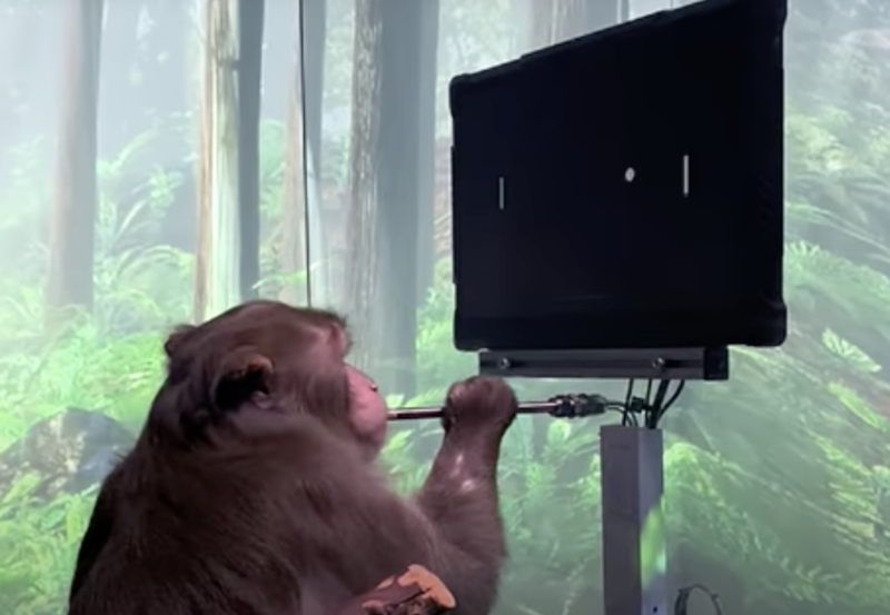
Enlarge / Pager, a 9-year-old Macaque, plays MindPong with his Neuralink. (credit: YouTube/NeuraLink)
The US Department of Transportation is investigating allegations that Elon Musk's brain-computer interface company, Neuralink, violated federal transportation regulations when it shipped contaminated implants removed from the brains of deceased research monkeys infected with multiple types of dangerous pathogens. The alleged violations could have put humans at risk of exposure to hazardous germs, including drug-resistant bacteria and a potentially life-threatening herpes virus.
Reuters was the first to report the department's investigation, which was sparked by allegations brought Thursday by the Physicians Committee for Responsible Medicine (PCRM), a medical group that advocates for animal welfare in medical research. The Department of Transportation confirmed to Ars on Friday that it has opened a standard investigation of Neuralink in response to PCRM's allegations.
In a letter addressed to Transportation Secretary Pete Buttigieg and William Schoonover, associate administrator of the department's Pipeline and Hazardous Material Safety Administration, the PCRM laid out its evidence for possible violations of hazardous material transportation regulations based on a trove of documents and emails obtained through public record requests. The advocacy group says the evidence shows Neuralink's contaminated hardware was not properly packaged to prevent exposure to humans and that Neuralink employees who transported the material had failed to undergo legally required training on how to safely transport such material.
A 319-million-year-old fossilized fish skull holds the oldest example of a well-preserved vertebrate brain.
Scientists pulled the skull from a coal mine in England more than a century ago. The brain and its cranial nerves are roughly an inch long and belong to an extinct bluegill-size fish. The discovery opens a window into the neural anatomy and early evolution of the major group of fishes alive today, the ray-finned fishes, according to the study in Nature.
The serendipitous find also provides insights into the preservation of soft parts in fossils of backboned animals. Most of the animal fossils in museum collections were formed from hard body parts such as bones, teeth, and shells.
The CT-scanned brain analyzed for the new study belongs to Coccocephalus wildi, an early ray-finned fish that swam in an estuary and likely dined on small crustaceans, aquatic insects, and cephalopods, a group that today includes squid, octopuses, and cuttlefish. Ray-finned fishes have backbones and fins supported by bony rods called rays.
When the fish died, the soft tissues of its brain and cranial nerves were replaced during the fossilization process with a dense mineral that preserved, in exquisite detail, their three-dimensional structure.
“An important conclusion is that these kinds of soft parts can be preserved, and they may be preserved in fossils that we’ve had for a long time—this is a fossil that’s been known for over 100 years,” says senior author Matt Friedman, a paleontologist and director of the Museum of Paleontology at the University of Michigan.
“Not only does this superficially unimpressive and small fossil show us the oldest example of a fossilized vertebrate brain, but it also shows that much of what we thought about brain evolution from living species alone will need reworking,” says lead author Rodrigo Figueroa, a doctoral student who did the work as part of his dissertation, under Friedman, in the earth and environmental sciences department.
“With the widespread availability of modern imaging techniques, I would not be surprised if we find that fossil brains and other soft parts are much more common than we previously thought. From now on, our research group and others will look at fossil fish heads with a new and different perspective.”
The skull fossil from England is the only known specimen of its species, so only nondestructive techniques could be used during the study.
The work on Coccocephalus is part of a broader effort by Friedman, Figueroa, and colleagues that uses computed tomography (CT) scanning to peer inside the skulls of early ray-finned fishes. The goal of the larger study is to obtain internal anatomical details that provide insights about evolutionary relationships.
In the case of C. wildi, Friedman wasn’t looking for a brain when he fired up his micro-CT scanner and examined the skull fossil.
“I scanned it, then I loaded the data into the software we use to visualize these scans and noticed that there was an unusual, distinct object inside the skull,” he says.
The unidentified blob was brighter on the CT image—and therefore likely denser—than the bones of the skull or the surrounding rock.
“It is common to see amorphous mineral growths in fossils, but this object had a clearly defined structure,” Friedman says.
The mystery object displayed several features found in vertebrate brains: It was bilaterally symmetrical, it contained hollow spaces similar in appearance to ventricles, and it had multiple filaments extending toward openings in the braincase, similar in appearance to cranial nerves, which travel through such canals in living species.
“It had all these features, and I said to myself, ‘Is this really a brain that I’m looking at?'” Friedman says. “So I zoomed in on that region of the skull to make a second, higher-resolution scan, and it was very clear that that’s exactly what it had to be. And it was only because this was such an unambiguous example that we decided to take it further.”
Though preserved brain tissue has rarely been found in vertebrate fossils, scientists have had better success with invertebrates. For example, the intact brain of a 310-million-year-old horseshoe crab was reported in 2021, and scans of amber-encased insects have revealed brains and other organs. There is even evidence of brains and other parts of the nervous system recorded in flattened specimens more than 500 million years old.
The preserved brain of a 300-million-year-old shark relative was reported in 2009. But sharks, rays, and skates are cartilaginous fishes, which today hold relatively few species compared to the ray-finned fish lineage containing Coccocephalus.
Early ray-finned fishes like Coccocephalus can tell scientists about the initial evolutionary phases of today’s most diverse fish group, which includes everything from trout to tuna, seahorses to flounder.
There are roughly 30,000 ray-finned fish species, and they account for about half of all backboned animal species. The other half is split between land vertebrates—birds, mammals, reptiles, and amphibians—and less diverse fish groups like jawless fishes and cartilaginous fishes.
The Coccocephalus skull fossil is on loan to Friedman from England’s Manchester Museum. It was recovered from the roof of the Mountain Fourfoot coal mine in Lancashire and was first scientifically described in 1925. The fossil was found in a layer of soapstone adjacent to a coal seam in the mine.
Though only its skull was recovered, scientists believe that C. wildi would have been 6 to 8 inches long. Judging from its jaw shape and its teeth, it was probably a carnivore, Figueroa says.
When the fish died, scientists suspect it was quickly buried in sediments with little oxygen present. Such environments can slow the decomposition of soft body parts.
In addition, a chemical micro-environment inside the skull’s braincase may have helped to preserve the delicate brain tissues and to replace them with a dense mineral, possibly pyrite, Figueroa says.
Evidence supporting this idea comes from the cranial nerves, which send electrical signals between the brain and the sensory organs. In the Coccocephalus fossil, the cranial nerves are intact inside the braincase but disappear as they exit the skull.
“There seems to be, inside this tightly enclosed void in the skull, a little micro-environment that is conducive to the replacement of those soft parts with some kind of mineral phase, capturing the shape of tissues that would otherwise simply decay away,” Friedman says.
Detailed analysis of the fossil, along with comparisons to the brains of modern-fish specimens from the University of Michigan Museum of Zoology collection, revealed that the brain of Coccocephalus has a raisin-size central body with three main regions that roughly correspond to the forebrain, midbrain, and hindbrain in living fishes.
Cranial nerves project from both sides of the central body. Viewed as a single unit, the central body and the cranial nerves resemble a tiny crustacean, such as a lobster or a crab, with projecting arms, legs and claws.
Notably, the brain structure of Coccocephalus indicates a more complicated pattern of fish-brain evolution than is suggested by living species alone, according to the authors.
“These features give the fossil real value in understanding patterns of brain evolution, rather than simply being a curiosity of unexpected preservation,” Figueroa says.
For example, all living ray-finned fishes have an everted brain, meaning that the brains of embryonic fish develop by folding tissues from the inside of the embryo outward, like a sock turned inside out.
All other vertebrates have evaginated brains, meaning that neural tissue in developing brains folds inward.
“Unlike all living ray-finned fishes, the brain of Coccocephalus folds inward,” Friedman says. “So, this fossil is capturing a time before that signature feature of ray-finned fish brains evolved. This provides us with some constraints on when this trait evolved—something that we did not have a good handle on before the new data on Coccocephalus.”
Comparisons to living fishes showed that the brain of Coccocephalus is most similar to the brains of sturgeons and paddlefish, which are often called “primitive” fishes because they diverged from all other living ray-finned fishes more than 300 million years ago.
Friedman and Figueroa are continuing to CT scan the skulls of ray-finned fish fossils, including several specimens that Figueroa brought to Ann Arbor on loan from institutions in his home country, Brazil. Figueroa says his doctoral dissertation was delayed by the COVID-19 pandemic but is expected to be completed in summer 2024.
Friedman and Figueroa says the discovery highlights the importance of preserving specimens in paleontology and zoology museums.
“Here we’ve found remarkable preservation in a fossil examined several times before by multiple people over the past century,” Friedman says. “But because we have these new tools for looking inside of fossils, it reveals another layer of information to us.
“That’s why holding onto the physical specimens is so important. Because who knows, in 100 years, what people might be able to do with the fossils in our collections now.”
The study includes data produced at University of Michigan’s Computed Tomography in Earth and Environmental Science facility, which is supported by the Department of Earth and Environmental Sciences and the College of Literature, Science, and the Arts.
Sam Giles of London’s Natural History Museum and the University of Birmingham is a senior author of the study. Additional coauthors are from the University of Chicago and the University of Michigan Museum of Paleontology.
Source: University of Michigan
The post Super ancient fish skull holds oldest backboned animal brain fossil appeared first on Futurity.